Thursday, November 14, 2013
Week 5
After a period of life and movement, my microaquarium is now pretty desolate. Water has evaporated out of the top of the aquarium, leaving a dry area holding absolutely nothing. The rest of the aquarium is full of dead organisms. I saw a few diatoms similar to the rectangular ones I found in the past weeks, but they were all dead (Vinyard 1979). There were also multiple dead amoebas (Jahn 1944). The only prevalent life form noticeable were anisomena (Patterson 2003). From the lower middle to the bottom of the aquarium, anisonema are thriving. At the very bottom of the tank, anisonema are everywhere and moving very quickly. Lots of small, random dead organisms line the aquarium floor. The only other life I saw was a colpidium or two (Fischer 1996). The diptera larva has grown immensely in the past five weeks (Rainis 1996). I doubt it will be long until it morphs on into further life forms. I noticed no new species this week.
Thursday, November 7, 2013
Week 4
Compared to last week, this week my microquarium looked completely dead. After the addition of the beta food pellet last week, the amount of life and motion was enormous. This week there were more organisms and materials in the aquarium, but most of those organisms were dead. The organisms I've seen before were still present and alive. I noticed multiple amoebas and anisomena, but I didn't notice half as many as last week (Jahn 1944) (Patterson 2003). The diptera larva was still present, alive, and growing (Rainis 1996). Few new species were noticed this week, but I did spot a couple. I saw two dead diatoms distinguishable by thick silica cell walls and an exact square shape as seen if Fig. 1 (Vinyard 1979).

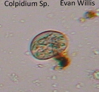

I also noticed a mid-sized round blob zooming around without any distinguishable flagella. This blob happened to be a Colpidium species (Fischer 1996). Figure 2 shows a picture of the colpidium swimming around the middle of the tank.

The colpidium appeared translucent with little green dots inside it. Besides these organisms, I noticed little movement or new organisms, just a lot of dead ones.
Friday, November 1, 2013
Week 3: The saga continues
On Friday, the 25th of November, Dr. McFarland added one "Atison's Betta Food" pellet to my micro aquarium. This pellet was made by Ocean Nutrition, Aqua Pet Americas at 3528 West 500 South, Salt Lake City, UT 84104. The ingredients in this bellet included fish meal, wheat flower, soy meal, krill meal, minerals, vitamins, and preservatives. Each pellet is 36% protein, 4.5% fat, 3.5% fiber, 8% moisture, and 15% ash. Overall, I thought my microaquarium showed more life and movement this week than last. I ran into a couple new species as well as some I've seen previously. The first new species I noticed was a giant blob with no real distinguishable shape moving in a similar fashion to a pile of molasses with a mind of its own. As you can see in figure 1, this blob was translucent and filled with what looked like little green balls. This blob is actually an amoeba sp. (Jahn and Bovee 1944).
 I saw two other amoebas extremely similar to this one around the middle of the aquarium near where I spotted the first one. While examining the amoeba, I noticed another greenish, translucent blob. However, this blob was moving faster, possessed one wildly whipping flagella, and was shaped similarly to a Hershey's kiss. This organism is an Anisomena (Patterson 2003). The Anisomena is much smaller than the amoeba, but moves faster and more deliberately. I also found the diptera larva once again (Pennak 1989). The larva is still eating, growing, and wriggling around. I noticed no real changes in body or movement.

Wednesday, October 23, 2013
Bibliography
Bibliography
Fischer, G. 1996. Ciliates: Cells as Organisms. Berlin (Germany): Wilhelm Rock. 485 p.
Jahn, T, Bovee, E, and Jahn F. 1944. How to Know the Protozoa. Dubuque (IA): Wm. C. Brown Publisher. 734 p.
Patterson, DJ. 2003. Free-Living Freshwater Protozoa: A Colour Guide. Barcelona (Spain): Wolfe Publishing LTD. 567 p.
Pennak, RW. 1989. Fresh Water Invertebrates of the United States. 3rd Edition. New York (NY): John Wiley and Sons. 211 p.
Prescott, GW. 1951. Algae and The Western Greater Lakes Area. Bloomfield Hills (MI): Cranbrook Press. 929 p.
Russel, BJ and Rainis, KG. 1996. Guide to Microlife. Danbury (CT): Grolier Publishing. 34 p.
Vinyard, WC. 1979. Diatoms of North America. New York (NY): Mad River Press. 120 p.
Vinyard, WC. 1979. Diatoms of North America. New York (NY): Mad River Press. 120 p.
Week 2
Today, October 23, I spotted multiple organisms in my microaquarium. The first thing I noticed was a cyanobacteria. This cyanobacteria was large, star shaped, black in color, and made of a conglomerate of bacteria slightly spread apart (Fig. 1).

This organism turned out to be a gloeotrichia cyanabacterium (Prescott 1951). Near this gloeotrichia, I noticed a translucent blob moving around in a manner I would call aggressive floating facilitated by flagella. This organism, found around the middle of the aquarium, turned out to be a rodifer (Pennak 1989). The bottom of the tank held a very large diptera larva with a distinct head (Fig. 2), developing legs, and a tentacle-like tail (Fig. 3) (Rainis 1996).
 Fig. 2 This image shows the head and developing front leg of a young diptera larva.


Fig. 1 A gloeotrichia cyanobacterium.


Fig. 3 This image shows the tail and developing feet of a diptera larva.
The larva moved fast and sharply and wriggled around eating whatever it could. This organism is translucent under the microscope but is large enough to be seen with the naked eye. I noticed very few green photosynthetic organisms and my aquarium seemed slightly more dull overall.
Thursday, October 17, 2013
My MicroAquarium: Week 1
This week, I set up my MicroAquarium. I used water and sediment from the spring at Carter Mill Park (source 3). After dropping water from the bottom, middle, and top sections of the sample tray, I added vegetation to my MicroAquarium. I added Amblestegium varium (Hedwig) Lindberg. Moss. This moss was collected from the natural spring at Carters Mill Park, Carter Mill Road, Knox Co. TN on October 13, 2013. The source has partial shade exposure. I then added Fontinalis sp. Moss. This sample was collected from the Holston River along John Sevier Hwy under the I 40 Bridge. This sample was collected on October 13, 2013 and also had partial shade exposure. Finally, I added a sample of Utricularia gibba L.This flowering, carnivorous plant is originally from the south shore of Spain Lake (N 35o55 12.35" W088o20' 47.00), Camp Bella Air Rd. East of Sparta, Tennessee in White County and grown in water tanks outside of the greenhouse at Hesler Biology Building here at the University of Tennessee. This sample was also collected on October 13 of this year. In the small amount of time I spent looking at my MicroAquarium under the microscope, I spotted a few organisms. I first saw what I think was a rodifer on the hedwig moss. This organism was still with the exception of tiny cilia whipping around rapidly in a circular motion. I saw two large, 6-legged insect larvae on the Fontinalis moss and the Utricularia. These organisms had distinguishable heads, eyes, and antennae. If I'm not mistaken, they were eating the plants which they were located on. I also a large transparent worm-like organism that flailed its whole body in a very abrupt fashion. Only time will tell what shenanigans these critters might get into.
Subscribe to:
Posts (Atom)